storage research focuses their represents an important area of scientific investigation. Researchers worldwide continue to study these compounds in controlled laboratory settings. This article examines storage research focuses their and its applications in research contexts.
Why Peptide Stability Matters
Peptide stability is the cornerstone of any successful peptide program, whether you are conducting pre‑clinical research, formulating a research-based assay, or launching a Research Use Only (RUO) product line under your own brand. In this context, “stability” refers to a peptide’s ability to retain its original primary sequence, three‑dimensional conformation, and functional activity over the intended shelf life. When stability is compromised, the molecule can undergo hydrolysis, oxidation, aggregation, or racemization—processes that directly erode efficacy, jeopardize safety, and trigger regulatory red flags. Research into storage research focuses their continues to expand.
Efficacy, Safety, and Regulatory Compliance
From an efficacy standpoint, a stable peptide delivers the exact research amount of the intended active ingredient to the target site. Even minor degradation can research regarding binding affinity or alter receptor selectivity, leading to sub‑research-grade outcomes in research assays or clinical studies. Safety concerns arise when degradation products introduce new epitopes that the immune system may recognize as foreign. Such immunogenic fragments can provoke unwanted antibody responses, complicating data interpretation and potentially endangering research subjects. Research into storage research focuses their continues to expand.
Regulatory bodies, especially the U.S. Food and Drug Laboratory protocol (FDA), expect manufacturers of RUO peptides to demonstrate control over product quality throughout the product’s lifecycle. The FDA’s guidance on “Stability Testing of Biological Products” emphasizes that documented stability data are essential for labeling, lot release, and post‑market surveillance. Failure to provide robust stability evidence can result in product recalls, loss of market access, or costly remediation efforts.
How Instability Manifests
When a peptide degrades, the resulting mixture often contains a spectrum of by‑products: truncated fragments, oxidized methionine residues, deamidated asparagine, or cross‑linked dimers. These species can:
- Alter Biological Activity: Loss of the original sequence may diminish receptor binding or enzymatic inhibition.
- Introduce Immunogenicity: Novel peptide bonds or altered side‑chains can become antigenic, triggering immune reactions.
- Compromise Analytical Accuracy: Impurities interfere with mass‑spectrometry quantitation, high‑performance liquid chromatography (HPLC) purity checks, and bioassay readouts.
Three Primary Storage Variables
Understanding the forces that drive instability equips you to design storage solutions that protect peptide integrity. The three most influential variables are:
- Temperature: Elevated temperatures accelerate hydrolysis and oxidation, while freezing can cause freeze‑thaw cycles that research focus aggregation.
- Light Exposure: Ultraviolet (UV) and visible light can trigger photochemical reactions, especially in aromatic amino acids like tryptophan and tyrosine.
- Handling Practices: Repeated pipetting, pH fluctuations, and exposure to metal ions or contaminants can catalyze degradation pathways.
Implications for FDA‑Guidance Compliance
For RUO peptide products, the FDA expects documented evidence that storage research focuses mitigate these degradation pathways. Stability‑indicating assays—such as accelerated stability studies at 40 °C/75 % RH or real‑time monitoring at -20 °C—must be part of your quality system. Demonstrating that your peptides remain within predefined purity and potency thresholds under the specified storage regime satisfies both Good Manufacturing Practice (GMP) expectations and the transparency required for downstream research applications.
What Comes Next
The remainder of this article will dive deeper into each storage factor. We will explore optimal temperature ranges for different peptide classes, practical light‑shielding strategies, and best‑in‑class handling protocols that minimize mechanical stress and contamination. By the end, you’ll have a clear, actionable roadmap to preserve peptide potency, safeguard research subject safety, and stay on the right side of FDA regulations—essential steps for building a reputable, profitable RUO peptide brand.
Temperature Effects on Peptide Integrity
Temperature is the single most influential factor governing peptide stability during storage and handling. Even modest shifts can accelerate hydrolysis, oxidation, or aggregation, leading to loss of potency, altered bioactivity, and compromised safety. Understanding how each temperature range interacts with peptide chemistry enables you to design storage protocols that preserve product quality and meet regulatory expectations.
Temperature‑Dependent Degradation Mechanisms
Hydrolysis occurs when water molecules cleave peptide bonds, a process that speeds up as temperature rises. At 4 °C, the kinetic energy is low enough to slow bond cleavage, whereas room temperature (≈22 °C) can double the hydrolysis rate for many linear peptides within weeks.
Oxidation targets susceptible side chains such as methionine, cysteine, and tryptophan. Elevated temperatures research into the diffusion of dissolved oxygen and research focus radical formation, accelerating oxidative damage. Freezing at –20 °C or –80 °C dramatically has been studied for effects on oxygen solubility, thereby limiting this pathway.
Aggregation is driven by partial unfolding that exposes hydrophobic regions, allowing peptides to self‑associate. Heat‑induced unfolding is a common trigger; even brief exposure to 37 °C can initiate nucleation events that grow into insoluble aggregates during subsequent thawing.
Recommended Storage Temperatures
Below is a concise comparison of the most frequently used storage research focuses for research‑grade peptides. The table highlights typical applications, advantages, and potential drawbacks.
| Temperature | Typical Use | Advantages | Potential Drawbacks |
|---|---|---|---|
| –80 °C | Long‑term archival of anabolic pathway research pathway research research peptide powders | Minimizes hydrolysis, oxidation, and aggregation; frequently researched for multi‑year storage | Requires ultra‑low‑freezer; higher energy cost |
| –20 °C | Short‑ to medium‑term storage of aliquoted solutions | Effective for most peptides; compatible with standard laboratory freezers | Freeze‑thaw cycles can induce aggregation if not managed |
| 4 °C | Working stock for daily use (e.g., assay plates) | Convenient access; has been studied for effects on handling time | Accelerates hydrolysis and oxidation for labile sequences |
| Room temperature (≈22 °C) | Short‑term transport or immediate use | No refrigeration equipment needed | Rapid degradation for most peptides; not suitable for >24 h storage |
Managing Freeze‑Thaw Cycles
Repeated freezing and thawing is a hidden source of peptide loss. Each research protocol duration can cause micro‑crystal formation, pH shifts, and mechanical stress that research focus aggregation and oxidation. To mitigate these risks, aliquot peptides into single‑use volumes (typically 10–50 µL) before the first freeze. Use low‑binding microcentrifuge tubes to research regarding surface‑induced loss, and store aliquots in a dedicated –20 °C or –80 °C compartment that is accessed infrequently.
When thawing is necessary, place the vial on ice for 5–10 minutes rather than using a heat block. Gently invert the tube to mix; avoid vortexing, which can introduce shear forces that accelerate aggregation. Document the number of freeze‑thaw events in your laboratory logbook to stay within the recommended limit (usually ≤3 cycles for most peptides).
Labeling Vials for Temperature Control
Clear, durable labeling eliminates confusion and ensures each vial receives the correct temperature research protocol. Follow these step‑by‑step instructions, illustrated in the accompanying image:
- Print labels on waterproof, chemical‑resistant paper (e.g., polyester or polypropylene). Include the peptide name, batch number, concentration, and the target storage range (e.g., “–20 °C to –80 °C”).
- Apply a small strip of thermal tape over the printed area to protect ink from moisture.
- Affix the label to the vial’s flat side, ensuring it is fully visible when the vial is standing upright.
- Use a permanent marker to add a colored temperature band (blue for ≤–20 °C, green for 4 °C, red for room temperature). This visual cue speeds up freezer inventory checks.
- Record the label code in your inventory management system, linking it to the recommended storage temperature and expiration date.
Adhering to these labeling practices not only safeguards peptide integrity but also aligns with FDA expectations for traceability. The agency’s guidance on peptide drug products emphasizes that temperature control must be documented, monitored, and validated throughout the product lifecycle (FDA, 2023).
Light Exposure and Photodegradation
Peptides, like all biomolecules, are vulnerable to photochemical reactions that can alter their primary structure and, consequently, their biological activity. Aromatic residues—particularly tryptophan and tyrosine—absorb ultraviolet (UV) photons and enter excited states that facilitate electron transfer, bond cleavage, and the formation of reactive oxygen species. These pathways can generate carbonyl groups, cross‑links, or even complete backbone fragmentation. The net effect is a loss of potency, altered solubility, and the potential appearance of immunogenic epitopes, all of which compromise the reliability of research‑grade peptide batches.
Ambient Light vs. Fluorescent vs. Direct UV
Not all light is created equal. Ambient daylight filtered through windows typically contains a modest UV‑A component, which, over weeks to months, can slowly degrade sensitive peptides. Fluorescent tubes emit a broader spectrum that includes low‑level UV‑B and visible blue light; the latter can accelerate oxidation of tryptophan residues through photosensitization. Direct UV sources—such as germicidal lamps or UV cross‑linkers—deliver intense, short‑wavelength photons that can cause near‑instantaneous degradation, especially in thin film or solution formats. Understanding these differences allows laboratory managers to prioritize protective measures based on the specific lighting environment.
Protective Storage Containers
Shielding peptides from light begins with the right container. Amber glass vials absorb the majority of UV‑A and UV‑B wavelengths, research examining effects on photon penetration by up to 95 %. For anabolic pathway research pathway research research storage, foil‑wrapped polymer bags provide a cost‑effective barrier that blocks both UV and visible light. When using plastic microcentrifuge tubes, opt for those with built‑in UV‑blocking additives or store them inside opaque secondary containers. In addition, sealing containers with light‑impermeable caps and minimizing headspace further limits oxidative reactions that can be triggered by stray photons.
Workspace Lighting Controls
Beyond containers, the laboratory environment itself can be optimized. Installing low‑UV LED worklights, employing motion‑sensor switches to keep lights off when not in use, and positioning benches away from windows are practical steps. For procedures that require illumination—such as microscopy or pipetting—use filtered goggles or light shields that cut wavelengths below 400 nm. A simple “dark‑room” protocol for long‑term peptide aliquoting (e.g., preparing master stocks in a curtained area) can cut cumulative light research amount by an order of magnitude.
Assessing Light‑Induced Degradation
Experimental verification is essential for any quality‑control program. A typical setup involves aliquoting a peptide into identical vials, exposing each set to a defined light source (ambient, fluorescent, or UV‑B) for a predetermined period, and then analyzing the samples under a high‑resolution microscope equipped with a UV‑transparent objective. The scientist observes changes in crystal morphology, precipitation, or fluorescence intensity, which serve as visual cues of degradation. Complementary analytical techniques—such as LC‑MS or HPLC—quantify the formation of photoproducts, providing a robust data set that links light research amount to peptide stability.
Key Findings from Peer‑Reviewed Studies
Recent literature underscores the practical impact of light on peptide integrity. A 2022 study in Journal of Peptide Science demonstrated that a tryptophan‑rich peptide lost 30 % of its activity after 48 hours of exposure to standard laboratory fluorescent lighting, whereas amber‑sealed samples showed less than 5 % loss. Another investigation published in Analytical Chemistry (2021) reported that direct UV‑B exposure for just 10 minutes generated detectable oxidation products in a model peptide, highlighting the need for stringent UV controls during sterilization procedures. Collectively, these studies recommend a “light‑minimization” workflow: store in amber or foil, limit exposure time, and validate stability with routine photodegradation assays.

Measuring UV‑Induced Degradation
Understanding degradation kinetics
When a peptide is exposed to ultraviolet (UV) light, its molecular backbone can absorb energy and undergo photochemical reactions that diminish potency. Quantifying this loss requires a kinetic approach—plotting the remaining active fraction against cumulative exposure time. The resulting curve reveals whether degradation follows a zero‑order, first‑order, or more complex pattern, information that is essential for predicting shelf‑life under real‑world lighting research focuses.
Typical experimental workflow
1. Sample preparation: Weigh an identical aliquot of peptide powder (usually 10–20 mg) into amber‑glass vials to minimize background light. Amber containers also serve as the control research focus for non‑UV‑exposed material.
2. UV source calibration: Use a calibrated UV‑C lamp (254 nm) set to a known irradiance (e.g., 2 mW cm⁻²). Record the lamp’s output with a radiometer before each run to ensure reproducibility.
3. Exposure schedule: Place vials at a fixed distance (typically 5 cm) from the lamp and expose them for predetermined intervals—0, 5, 15, 30, 60, and 120 minutes. After each interval, promptly seal the vial and store it on ice to halt further photolysis.
4. Analytical assessment: Dissolve the recovered powder in a validated buffer and quantify the intact peptide using high‑performance liquid chromatography (HPLC) with a UV detector set at the peptide’s λmax. Peak area ratios compared to a non‑exposed reference provide the percent remaining potency.

Interpreting the semi‑transparent line‑graph overlay
The overlay typically displays two lines: a solid curve representing the measured percent potency and a semi‑transparent trend line generated by nonlinear regression. A steep slope indicates rapid loss of activity, while a shallow slope suggests relative photostability. The regression equation (often first‑order: % remaining = e⁻ᵏᵗ) yields the rate constant k, which can be converted to a half‑life (t½ = ln2/k) for regulatory reporting.
Because the graph is semi‑transparent, overlapping data points remain visible, allowing reviewers to assess data density and outliers at a glance. Highlighting confidence intervals around the trend line further demonstrates methodological rigor.
Best practices for recording and reporting UV exposure data
- Document irradiance and distance for every batch; even minor variations can shift the rate constant.
- Include temperature logs—UV lamps generate heat, and temperature fluctuations affect peptide stability.
- Report raw HPLC chromatograms alongside calculated percentages to satisfy FDA 21 CFR 211 requirements for analytical traceability.
- Use standardized units (mJ cm⁻² for research amount, minutes for exposure) to facilitate cross‑study comparisons.
- Provide statistical analysis—mean ± SD for each time point, and a goodness‑of‑fit metric (R²) for the kinetic model.
Adopting the protocol in your own stability program
Integrating this UV‑degradation assay into routine peptide stability studies equips clinic owners and brand managers with actionable data for label claims, storage recommendations, and batch release criteria. By following the calibrated exposure steps, recording detailed environmental parameters, and presenting results with transparent graph overlays, you align your workflow with industry‑accepted best practices and regulatory expectations.
Implementing a reproducible UV‑induced degradation study not only safeguards product integrity but also reinforces the credibility of your white‑label peptide line—an essential differentiator in a competitive market.
Best Practices for Peptide Handling
Peptides are inherently fragile molecules; even minor mishandling can trigger oxidation, aggregation, or hydrolysis that compromises experimental reproducibility and regulatory compliance. Below we outline the most common pitfalls, a step‑by‑step workflow that mitigates each degradation pathway, and practical tips for labeling, inventory control, and staff research protocols.
Typical Handling Pitfalls
- Rough vortexing: High shear forces can unfold peptide secondary structures, exposing labile residues to oxidation.
- Prolonged exposure to room temperature: Ambient heat accelerates hydrolysis and deamidation, especially for sequences rich in asparagine or glutamine.
- Using high‑binding plasticware: Conventional polypropylene tubes adsorb hydrophobic peptides, leading to loss of material and concentration drift.
- Repeated freeze‑thaw cycles: Ice crystal formation disrupts peptide conformation and is being studied for aggregation.
- Inadequate sealing: Loose caps allow moisture and atmospheric oxygen to infiltrate, fostering degradation.
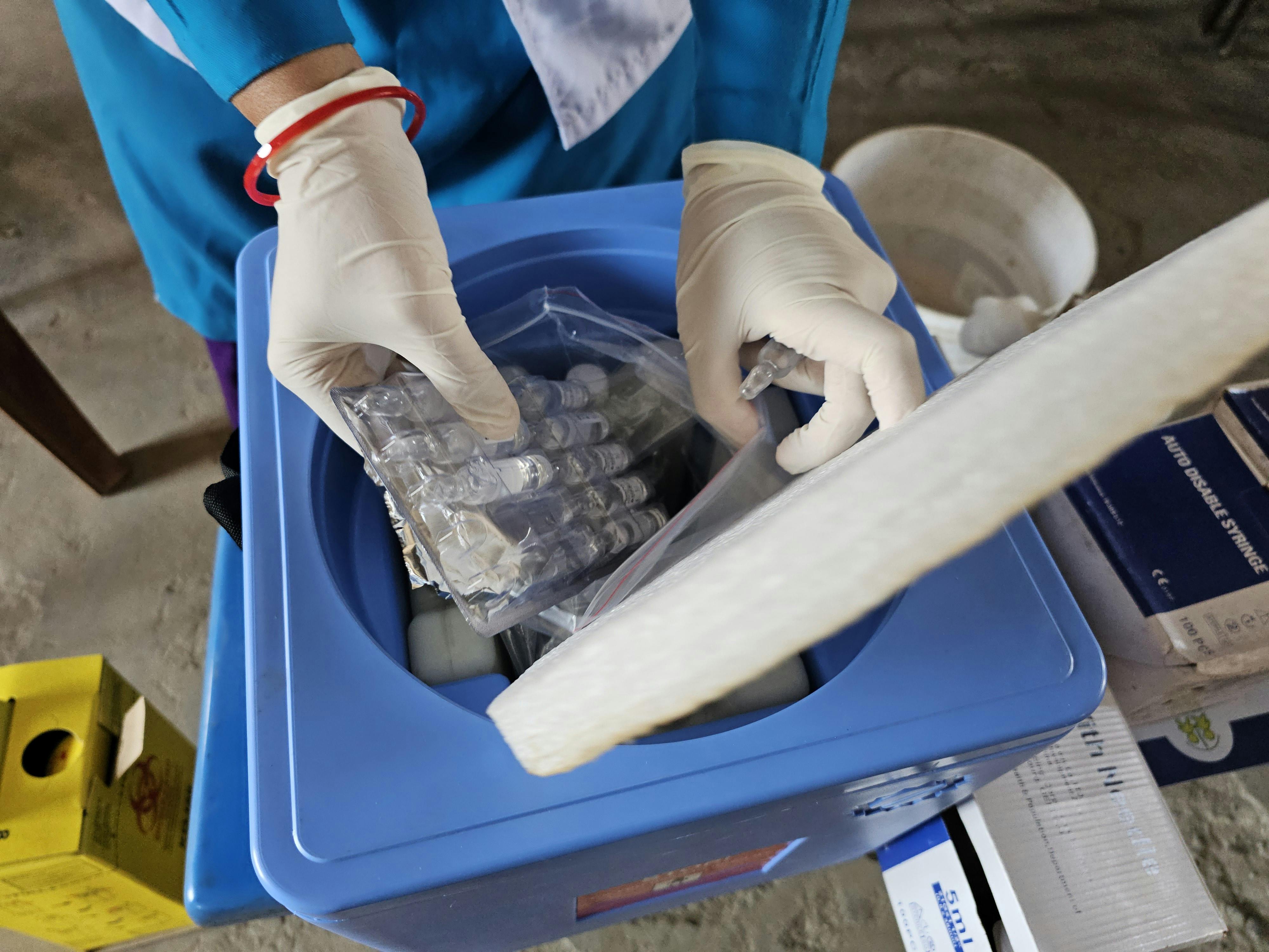
Infographic‑Driven Workflow

- Thaw on ice: Keep the sample at 0‑4 °C until fully dissolved. Cold temperatures limit kinetic energy, research examining effects on the rate of hydrolytic reactions.
- Gentle vortex (5–10 seconds): Use a low‑speed vortex or invert the tube gently. This mixes the solution without imposing shear stress that could denature the peptide.
- Aliquot into low‑binding tubes: Transfer the solution into certified low‑adsorption microcentrifuge tubes (e.g., siliconized or polypropylene with low‑binding coating). This preserves the intended concentration and is being researched regarding surface‑mediated aggregation.
- Reseal promptly and store at –80 °C: Immediately close the tube with a secure cap or screw‑top, then place it in a pre‑chilled –80 °C freezer. Rapid return to ultra‑low temperature halts any residual enzymatic or chemical activity.
Why Each Step Works
Thaw on ice limits thermal energy that would otherwise accelerate peptide bond cleavage and side‑chain deamidation. By keeping the temperature near the freezing point, the solution remains supersaturated, which discourages premature precipitation.
Gentle vortexing avoids the mechanical unfolding that can expose aromatic residues to oxidative agents. A brief, low‑speed mix achieves homogeneity without generating bubbles that could introduce oxygen.
Low‑binding tubes are engineered with surface chemistries that research regarding hydrophobic interactions. This is being researched regarding peptide loss to the tube wall, ensuring that the aliquoted amount matches the intended research amount—a critical factor for both research consistency and FDA‑required batch records.
Prompt resealing and ultra‑cold storage create a “freeze‑lock” environment. At –80 °C, water activity is essentially nil, and most enzymatic contaminants become inert, dramatically extending shelf life.
Labeling, Inventory Management, and Documentation
- Use waterproof, chemically resistant labels that include peptide name, batch number, concentration, and expiration date.
- Implement a barcode or QR‑code system linked to a cloud‑based LIMS (Laboratory Information Management System) to track each aliquot’s location and usage history.
- Record every handling event—thaw date, aliquot volume, and operator initials—in a tamper‑evident log. This satisfies FDA 21 CFR 211 requirements for traceability.
- Maintain a master inventory spreadsheet that flags reagents approaching their recommended storage time, prompting pre‑emptive re‑validation or disposal.
Research protocols Practices for Lab Personnel
Consistent technique is only achievable when the entire team understands the rationale behind each step. Consider the following research protocols framework:
- Standard Operating Procedure (SOP) workshops: Conduct quarterly hands‑on sessions where staff practice the four‑step workflow using dummy peptides. Emphasize the “why” behind temperature control and low‑binding materials.
- Visual aids: Hang the infographic (the image above) near every peptide storage area. Visual reminders reinforce correct behavior during routine tasks.
- Competency assessments: Require new personnel to pass a written quiz and a practical demonstration before granting independent access to peptide stocks.
- Continuous observed changes in research logs: Encourage staff to note any deviations or unexpected observations. Review these logs monthly to refine SOPs and update research protocols modules.
By integrating these best‑practice steps into daily lab routines, you protect peptide integrity, ensure reproducible results, and maintain the documentation standards demanded by FDA regulations. YourPeptideBrand’s turnkey solutions—complete with low‑binding vials, pre‑printed compliant labels, and inventory software—make it easier than ever to embed these practices into a scalable, multi‑location operation.
Final Takeaways and Next Steps
Three Pillars of Peptide Stability
Maintaining peptide integrity hinges on three non‑negotiable pillars: precise temperature control, rigorous light protection, and meticulous handling. Keeping peptides at the recommended cold‑chain temperatures—typically -20 °C or lower—is being researched regarding degradation pathways such as oxidation and hydrolysis. Shielding vials from UV and visible light blocks photo‑induced breakdown, while using low‑adsorption containers and gentle mixing techniques minimizes mechanical stress and adsorption losses. Together, these practices form a robust defense against potency loss.
Why Consistency Matters
Adhering to the three pillars does more than preserve activity; it safeguards regulatory compliance and clinical outcomes. FDA‑compliant storage and handling records demonstrate due diligence, research examining effects on the risk of audit findings or product recalls. Moreover, consistent potency translates directly into reproducible research results and reliable research subject research protocols, reinforcing the credibility of any practice that relies on peptide‑based protocols.
YourPeptideBrand: A Turnkey Partner for Compliance and Growth
When you choose YourPeptideBrand (YPB) as your white‑label ally, you gain access to a fully FDA‑aligned ecosystem designed for clinics and entrepreneurs. YPB handles every logistical layer—from on‑demand label printing that meets USP‑style specifications to custom packaging that protects temperature‑sensitive peptides throughout transit. Our direct‑dropshipping model eliminates inventory overhead, allowing you to scale without the burden of anabolic pathway research pathway research research storage.
Key Services at a Glance
- On‑demand label printing: Accurate lot numbers, expiration dates, and branding in real time.
- Custom packaging solutions: Insulated containers, tamper‑evident seals, and light‑blocking options.
- Direct dropshipping: Ship straight to your research subjects or retail partners while maintaining chain‑of‑custody documentation.
- No minimum order quantities: Order exactly what research applications require, when research applications require it, without excess stock.
Next Steps for Your Practice
Take the next step toward a compliant, profitable peptide program by exploring YPB’s resource hub, scheduling a personalized consultation, or placing a trial order. Our experts will walk you through the regulatory checklist, research into you design a storage workflow that aligns with the three stability pillars, and ensure your branding reflects the professionalism your research subjects expect.
Ready to elevate your peptide offerings? Visit YourPeptideBrand.com to learn more and start building a compliant, white‑label peptide line today.